
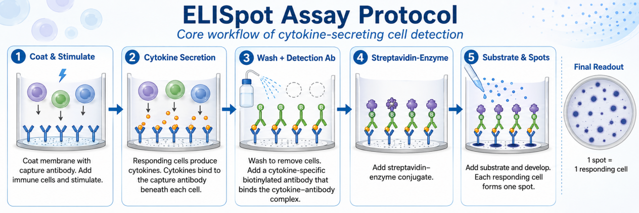
I. Experimental Principle
ELISpot (Enzyme-Linked Immunospot) is a highly sensitive assay for enumerating cells secreting target proteins (e.g., cytokines, antibodies) at the single-cell level. Effector cells are seeded on PVDF membrane plates coated with capture antibodies. Upon stimulation, secreted analytes are immobilized in the immediate vicinity of the secreting cell and detected through a cascade involving biotinylated detection antibodies, enzyme-linked streptavidin, and insoluble chromogenic substrates. Each discrete spot represents a single antigen-specific or cytokine-secreting cell, enabling direct quantification of cellular immune responses.
Unlike ELISA, which detects the total amount of free cytokines in body fluids, ELISpot detects the number of functionally secreting cells.
II. Materials and Reagents
III. Procedures
Stage 1: Plate Preprocessing and Coating (Afternoon of Day 0)
Stage 2: Cell Preparation (Day 1)
PBMC Isolation (Fresh Samples):
- Dilute whole blood with an equal volume of sterile normal saline or PBS
- Carefully overlay onto an equal volume of Ficoll-Paque without mixing
- Centrifuge at 700×g for 20 minutes at room temperature, brake off
- Aspirate the PBMC interface layer and wash twice with PBS (600×g, 5 min, room temperature)
- Count and adjust cell density
Cell Thawing (Cryopreserved Samples):
- Thaw quickly in a 37°C water bath
- Transfer to a 50 mL tube containing 15 mL of medium and mix gently
- Centrifuge at 300×g for 5 minutes
- Discard the supernatant, resuspend and count cells, then adjust the density
Cell Resting (Recommended for Cryopreserved Samples):
After thawing, rest for 3-6 hours (R10 (RPMI-1640 + 10% FBS), 37°C, 5% CO₂) to recover function
Stage 3: Cell Incubation and Stimulation (Day 1-2)
Stage 4: Detection Antibody Incubation (Day 2)
Stage 5: Enzyme Labeling and Chromogenic Reaction (Day 2)
IV. Data Analysis
Spot Counting: Use an ELISpot reader (AID, ImmunoSpot, CTL, etc.) or manual microscopy for counting
Parameter Setting: Define parameters such as spot size, intensity, and roundness, and keep the parameters consistent in the same experiment
Result Calculation: Spot-Forming Cells per 10⁶ cells (SFC/10⁶) = (spots/well) / (cells/well) × 10⁶
Plate Storage: Dried plates can be stored at room temperature in the dark for up to 3 months
V. Control Settings
VI. Key Notes
Ethanol activation strictly follows plate type parameters: MSIP (35% ≤1min) vs MAIPSWU (70% ≤2min); over time will damage the membrane
Cell Viability: Recommended >90%; dead cells cause high background and false positives
Do not move the plate during incubation: Prevent "snail trail" tailing spots
Washing Method: Both manual and automatic are acceptable; automatic plate washer needs to lower the flow rate to avoid membrane damage
Serum Selection: Use serum-free medium as much as possible, or screen low endotoxin serum
Antibody Filtration: 0.2 μm filtration of detection antibodies and enzyme-labeled reagents can reduce background
AEC Substrate: Suspected carcinogen; wear gloves during operation