
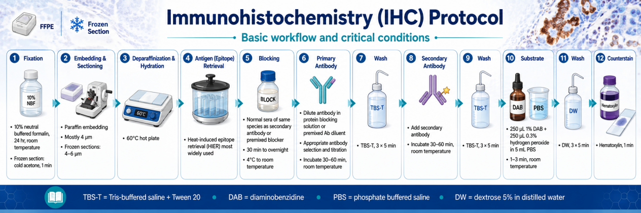
1. Reagents and Materials
2. Tissue Preparation
2.1 FFPE Tissue Sections
Fixation
- Immerse tissue in 10% NBF for 6-24 h at room temperature.
- Avoid over-fixation (>48 h) which masks epitopes; under-fixation (<30 min) causes antigen diffusion.
Dehydration and Clearing
- Dehydrate through graded ethanol: 50% → 70% → 80% → 95% → 100% (30 min each).
- Clear in xylene: 2-3 changes, 20 min each.
Infiltration and Embedding
- Infiltrate with paraffin at ≤60 °C: 3 changes, 1 h each.
- Embed in block; orient tissue as desired. Store at room temperature.
Sectioning
- Cut 4-5 μm sections on a microtome.
- Float sections on a 40-45 °C water bath; mount on silane-coated or gelatin-coated slides.
- Dry slides overnight at room temperature or 37 °C oven.
2.2 Frozen Tissue Sections
Snap-Freezing
- Freeze tissue in OCT compound on dry ice or in liquid nitrogen.
- Store at −80 °C.
Cryosectioning
- Equilibrate cryostat to −15 to −23 °C.
- Cut 5-10 μm sections; mount on charged slides.
- Air-dry 10-15 min.
Fixation
- Fix in cold acetone (100%) for 1 min, or 3.7% PFA for 4-24 h at RT.
- Wash in PBS.
3. Deparaffinization and Rehydration (FFPE only)
Do not allow slides to dry from this point forward. Drying causes non-specific antibody binding and high background.
4. Antigen Retrieval (AR)
4.1 Heat-Induced Epitope Retrieval (HIER) — Most Common
Methods:
- Water bath: Immerse slides in buffer-filled Coplin jar; heat to 95-99 °C.
- Pressure cooker / autoclave: 120 °C, 10 min.
- Microwave: 750-800 W, 10 min; monitor buffer level to prevent evaporation.
Post-HIER: Cool slides to room temperature in buffer (~15-20 min); rinse in distilled water.
4.2 Protease-Induced Epitope Retrieval (PIER)
Terminate enzymatic reaction by washing in PBS. PIER is harder to control and may damage tissue morphology.
5. Blocking and Quenching
Endogenous Peroxidase Quenching (chromogenic HRP systems only)
- Incubate in 0.3-3% H₂O₂ in methanol or PBS for 10-30 min at RT.
- Wash 2 × 5 min in PBS/TBS.
Protein Blocking
- Apply 2-10% normal serum from the secondary antibody host species in PBS/TBS.
- Incubate 30-60 min at RT in a humidified chamber.
- Alternatively: 1% BSA or commercial blocking buffer.
Avidin/Biotin Blocking (if using biotin-avidin amplification)
- Block endogenous biotin with avidin/biotin blocking kit.
6. Antibody Incubation
6.1 Primary Antibody
- Dilution: 1:50-1:500 (verify by datasheet; titrate empirically)
- Diluent: PBS/TBS with 1% BSA or normal serum
- Incubation: Overnight at 4 °C (optimal specificity) or 1-2 h at RT
- Volume: Sufficient to cover tissue section (~100-250 μL per section)
Include negative controls (omit primary antibody; replace with isotype-matched IgG or serum).
6.2 Washing
Wash 3 × 5 min with PBS-T or TBS-T at RT.
6.3 Secondary Antibody
- Conjugate: HRP, AP, or fluorophore (e.g., Alexa Fluor, Dylight)
- Dilution: 1:200-1:500 (HRP); 1:500-1:1,000 (fluorescent)
- Incubation: 30-60 min at RT in humidified chamber, protected from light (fluorescent)
6.4 Final Washing
Wash 3 × 5 min with PBS-T/TBS-T.
7. Detection
7.1 Chromogenic Detection (HRP-DAB)
- Prepare DAB substrate: mix DAB (1 mg/mL, freshly prepared) with 0.03% H₂O₂ in PBS.
- Apply to tissue; incubate 1-3 min at RT until brown precipitate develops.
- Monitor under microscope; stop reaction by rinsing in distilled water.
- Counterstain: Hematoxylin, 1-5 min; 1% HCl in 70% ethanol; blue in tap water.
- Dehydrate through ethanol series; clear in xylene; mount with permanent medium.
7.2 Fluorescent Detection
- Apply fluorophore-conjugated secondary antibody (as above).
- Counterstain nuclei with DAPI (1 μg/mL) or Hoechst 33342 for 5 min.
- Wash in PBS.
- Mount with antifade mounting medium.
- Image immediately or store at 4 °C, protected from light.
8. Multiplex IHC (Optional)
- Use primary antibodies from different species or isotype-specific secondaries.
- Sequential staining: complete first round (primary → secondary → detection), then strip or block, and repeat.
- For fluorescent multiplex: select spectrally distinct fluorophores; verify minimal spectral overlap.
9. Data Quantification
- IHC data are semi-quantitative.
- Score staining intensity (0-3) and percentage of positive cells (0-100%).
- Report as H-score or Allred score for clinical samples.
- Use image analysis software (e.g., ImageJ, QuPath) for objective quantification.
10. Critical Quality Control & Troubleshooting
11. Safety and Waste Disposal
- Handle xylene, DAB, and formaldehyde in a chemical fume hood. DAB is carcinogenic.
- Dispose of DAB-containing reagents and xylene per institutional hazardous waste protocols.
- Wear nitrile gloves and lab coat throughout procedure.
References
- Shi SR, Liu C, Taylor CR. Standardization of immunohistochemistry for formalin-fixed, paraffin-embedded tissue sections based on the antigen-retrieval technique. J Histochem Cytochem. 2007;55(2):105-9.
- Kim KM, Kwon MS, Park HS. Immunohistochemistry for Pathologists: Protocols, Pitfalls, and Tips. J Pathol Transl Med. 2016;50(6):411-8.
- Thermo Fisher Scientific. IHC Troubleshooting Guide.
- R&D Systems. Troubleshooting Guide: Immunohistochemistry.