Home/Western Blot Protocol 1. Reagents and Materials
2. Sample Preparation
1. Cell Lysis
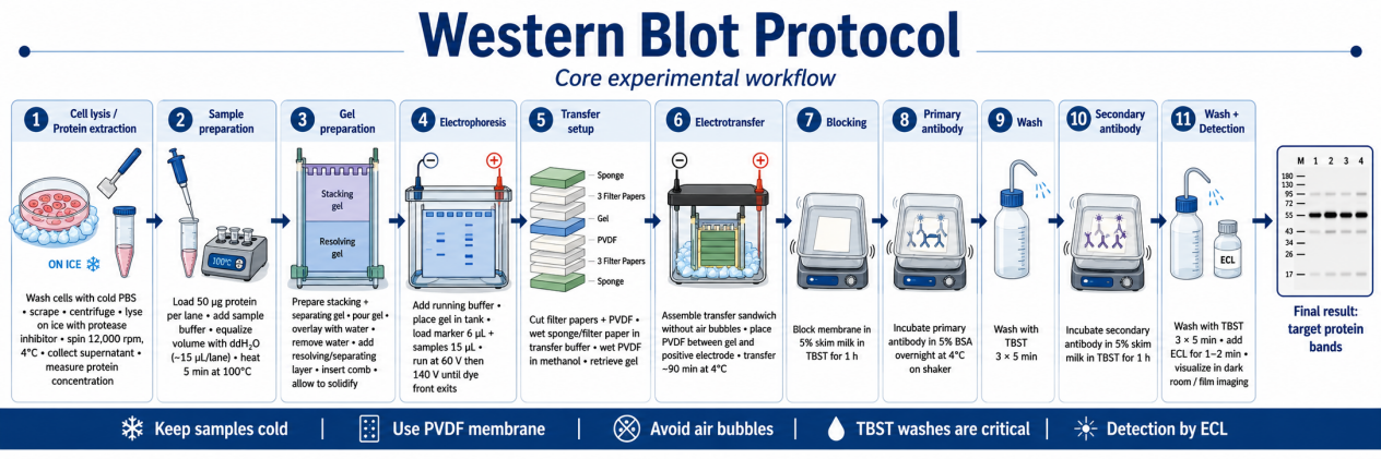
- Wash adherent cells 2× with ice-cold PBS; suspend cells in pre-chilled lysis buffer (e.g., RIPA with inhibitors).
- Incubate on ice for 15-30 min with occasional vortexing.
- Clarify lysate by centrifugation at 12,000-20,000 × g, 4 °C, 10-15 min.
- Collect supernatant; avoid disturbing the pellet.
2. Tissue Homogenization
- Snap-freeze tissue in liquid nitrogen; grind to powder.
- Homogenize in lysis buffer using a mechanical homogenizer or sonicator (pulse on ice).
- Clarify as above.
3. Protein Quantification
- Perform BCA assay according to manufacturer instructions.
- Prepare equal protein amounts (typically 20-40 μg per lane) in a final volume of 15-25 μL.
4. Denaturation
- Mix protein samples with 4×-6× SDS loading buffer containing reducing agent.
- Heat at 95 °C for 5-10 min (cytosolic and nuclear proteins). For membrane proteins, use 37 °C for 30 min to reduce aggregation.
- Briefly spin to collect condensation; load immediately or store at −20 °C.
3. SDS-PAGE
1. Gel Selection
- 8% gel: proteins >100 kDa
- 10-12% gel: proteins 20-100 kDa
- 15% gel: proteins <20 kDa (e.g., histones)
2. Assembly and Loading
- Assemble gel in electrophoresis tank; fill with 1× running buffer.
- Load protein samples and prestained molecular weight marker.
- Run at 80 V through the stacking gel (approx. 30 min), then increase to 120-200 V through the resolving gel until the dye front reaches the bottom.
4. Protein Transfer
1. Membrane Preparation
- PVDF: Immerse in 100% methanol for 30 s, then equilibrate in transfer buffer.
- Nitrocellulose: Equilibrate directly in transfer buffer.
2. Transfer Setup
- Assemble transfer sandwich: anode → filter paper → membrane → gel → filter paper → cathode.
- Roll out air bubbles meticulously using a glass rod or pipette; trapped bubbles cause patchy transfer.
4. Transfer Validation
- Stain membrane with Ponceau S (reversible) or stain the gel post-transfer with Coomassie to confirm complete transfer.
5. Blocking
- Incubate membrane in blocking buffer:
- - 5% non-fat dry milk in TBST (general use), or
- - 3-5% BSA in TBST (phospho-proteins, biotin systems)
- Duration: 1 h at room temperature with gentle rocking, or overnight at 4 °C for sensitive targets.
- Wash 3 × 5 min with TBST before antibody incubation.
6. Antibody Incubation
1. Primary Antibody
- Dilution: 1:500-1:5,000 (typically 1:1,000; titrate based on signal intensity)
- Diluent: Blocking buffer or commercial antibody diluent
- Incubation: Overnight at 4 °C with gentle agitation for optimal specificity
2. Washing
- Wash membrane 3 × 5-10 min with TBST at room temperature.
3. Secondary Antibody
- Dilution: 1:5,000-1:10,000
- Diluent: Blocking buffer or TBST
- Incubation: 1 h at room temperature with gentle rocking
4. Final Washing
- Wash membrane 3 × 5-10 min with TBST to remove unbound secondary antibody.
7. Signal Detection
1. Chemiluminescence (ECL)
- Mix ECL substrate (peroxide and luminol solutions) 1:1.
- Incubate membrane for 1-5 min; drain excess reagent.
- Capture image using chemiluminescence imager or X-ray film.
- Perform multiple exposures (short to long) to avoid signal saturation.
2. Fluorescence Detection
- Use IRDye-conjugated secondary antibodies.
- Image on a fluorescence scanner.
8. Stripping and Re-probing (Optional)
- Incubate membrane with stripping buffer (e.g., mild glycine-based or commercial buffer) for 10-20 min at room temperature with agitation.
- Wash extensively with TBST.
- Validate stripping efficiency by exposing to ECL without antibody; no signal should appear.
- Re-block and probe with new primary antibody (e.g., loading control).
9. Data Quantification and Normalization
- Western blot data are semi-quantitative.
- Normalize target band intensity to a housekeeping protein (GAPDH, α-Tubulin, β-Actin).
- Ensure signal intensity falls within the linear dynamic range of detection; avoid overexposure.
- Report relative fold change compared to control group.
10. Critical Quality Control & Troubleshooting
11. Safety and Waste Disposal
- Handle acrylamide, methanol, and β-mercaptoethanol in a chemical fume hood.
- Dispose of acrylamide gels and methanol-containing buffers per institutional hazardous waste protocols.
- Wear nitrile gloves when handling membranes and ECL reagents.
References
- Mahmood T, Yang PC. Western blot: technique, theory, and trouble shooting. N Am J Med Sci. 2012 Sep;4(9):429-34. doi: 10.4103/1947-2714.100998. PMID: 23050259; PMCID: PMC3456489.